
The Chemistry of Living Things
A Guide for IMAT Preparation
I. Introduction: The Chemical Basis of Life
Living organisms are composed of a unique and complex array of molecules, collectively known as biomolecules. These are primarily carbon-based compounds, and...
II. Carbohydrates: Energy and Structure
Carbohydrates are a major class of biomolecules with the general empirical formula , often called saccharides or sugars. They serve as energy sources and structural components in living organisms.
A. Monosaccharides: The Simplest Sugars
Monosaccharides are the fundamental building blocks of carbohydrates; they are simple sugars that cannot be broken down into smaller carbohydrate units by...
Glucose () is a particularly important hexose, serving as a primary energy source for cells...
Monosaccharides like glucose, galactose, and fructose are isomers, meaning they share the same chemical formula ()...
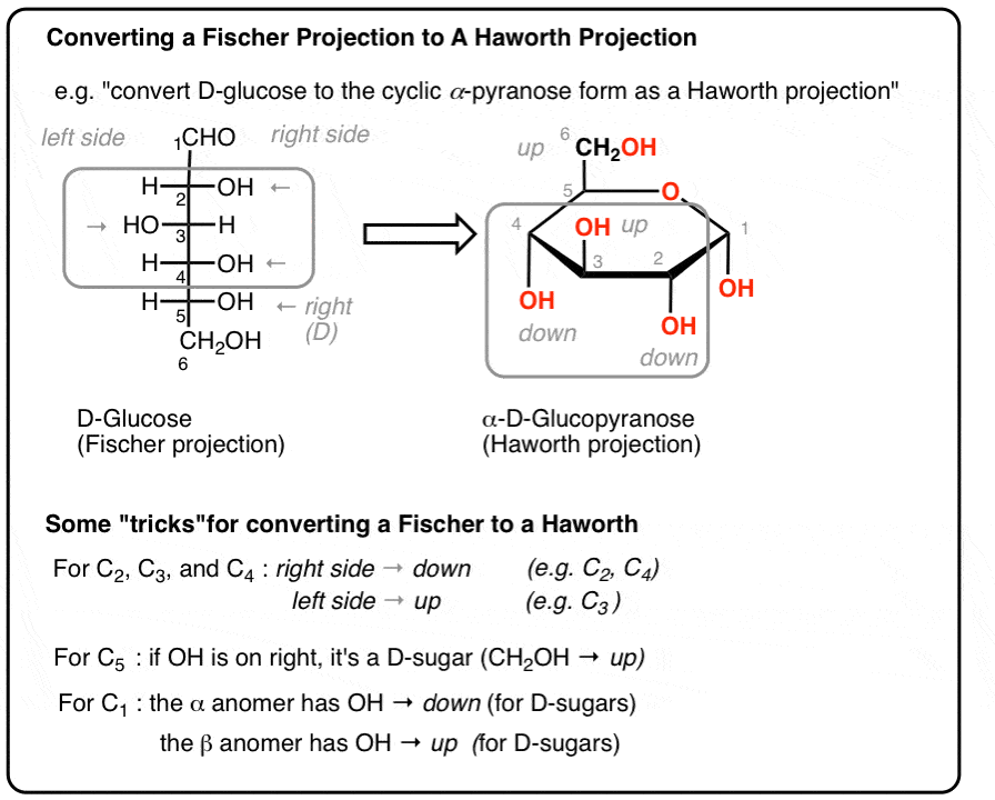
📸 Source/Description: Conversion between Fischer and Haworth projections
B. Disaccharides: Double Sugars
Disaccharides are formed when two monosaccharides are joined together by a glycosidic bond. This bond formation is a dehydration synthesis (or condensation)...
- Sucrose (common table sugar): Composed of glucose and fructose linked by an α-1,β-2 glycosidic bond.
- Lactose (milk sugar): Composed of glucose and galactose.
- Maltose (malt sugar): Composed of two glucose units.
Disaccharides serve as readily available energy sources. For instance, sucrose is a major transport form of sugar in plants. To be utilized by the body, disaccharides...
The nature of the glycosidic bond in a disaccharide significantly influences its chemical properties. For example, in sucrose, the glycosidic bond involves...

📸 Source/Description: Structure of sucrose molecule
C. Polysaccharides: Complex Carbohydrates
Polysaccharides are polymers composed of many monosaccharide units (often hundreds or thousands) linked by glycosidic bonds. They can be linear or branched...
Starch
Starch is the primary storage polysaccharide in plants, composed entirely of glucose units. It exists in two forms:
- Amylose: A linear, unbranched polymer where glucose units are linked by α-1,4 glycosidic bonds.
- Amylopectin: A branched polymer with a backbone of α-1,4 linked glucose units and branch points formed by α-1,6 glycosidic bonds.
Starch serves as a major energy reserve in plants and a crucial dietary source of energy for humans.

📸 Source/Description: Structure of amylose and amylopectin
Glycogen
Glycogen is the main storage polysaccharide in animals, often referred to as "animal starch". It is a polymer of glucose, structurally similar to amylopectin but more highly branched.

📸 Source/Description: Skeletal formula of glycogen
Cellulose
Cellulose is the most abundant structural polysaccharide in nature, forming the primary component of plant cell walls. It is an unbranched, linear polymer...

📸 Source/Description: Chemical structure of cellulose
| Table 1: Comparison of Key Polysaccharides | |||
|---|---|---|---|
| Feature | Starch | Glycogen | Cellulose |
| Monomer Unit | α-glucose | α-glucose | β-glucose |
| Glycosidic Linkage(s) | α-1,4 and α-1,6 | α-1,4 and α-1,6 | β-1,4 |
| Branching | Moderately branched | Highly branched | Unbranched (Linear) |
| Primary Function | Energy storage in plants | Energy storage in animals | Structural support in plants |
| Digestibility by Humans | Yes | (Synthesized in body) | No (Dietary fiber) |
III. Lipids: Diverse Roles in Energy, Membranes, and Signaling
Lipids are a diverse group of hydrophobic (water-fearing) biomolecules that are soluble in organic solvents but largely insoluble in water. They play crucial roles in energy storage, membrane structure, and signaling.
A. Triglycerides (Neutral Fats)
Triglycerides are esters formed from one molecule of glycerol and three fatty acid molecules. Fatty acids are long hydrocarbon chains with a terminal carboxyl group.

📸 Source/Description: Shorthand formula of triglyceride
B. Phospholipids
Phospholipids are structurally similar to triglycerides, but one fatty acid is replaced by a phosphate group, making the molecule amphipathic (having both hydrophilic and hydrophobic regions).

📸 Source/Description: Phospholipid bilayer in cell membrane
C. Steroids
Steroids are lipids characterized by a distinctive four-ring carbon skeleton. Cholesterol is a vital steroid in animals, acting as an essential component of cell membranes and precursor for steroid hormones.

📸 Source/Description: Structure of steroid backbone
| Table 2: Overview of Key Lipid Types | ||
|---|---|---|
| Lipid Type | Key Structural Features | Primary Function(s) |
| Triglycerides | Glycerol + 3 Fatty Acids | Long-term energy storage, insulation |
| Phospholipids | Glycerol + 2 Fatty Acids + Phosphate group (Amphipathic) | Main structural component of cell membranes |
| Steroids | Four-fused carbon ring structure | Membrane fluidity, signaling (hormones) |
IV. Proteins: The Workhorses of the Cell
A. Amino Acids: The Building Blocks
Proteins are polymers made up of monomeric units called amino acids. There are 20 common amino acids found in proteins. Each amino acid has a central carbon atom (α-carbon) bonded to an amino group, a carboxyl group, a hydrogen atom, and a variable R group.

📸 Source/Description: General structure showing R group
B. Peptide Bonds: Linking Amino Acids
Amino acids are linked together by peptide bonds to form polypeptide chains. A peptide bond is an amide linkage formed between the carboxyl group of one amino acid and the amino group of another.

📸 Source/Description: Peptide bond formation (condensation reaction)
C. Levels of Protein Structure
The functionality of a protein is intimately linked to its three-dimensional structure, organized hierarchically into four levels:
- Primary Structure (1°): The unique linear sequence of amino acids in a polypeptide chain, held together by peptide bonds.
- Secondary Structure (2°): Local, regular folding patterns of the polypeptide backbone, stabilized by hydrogen bonds. The two most common types are α-helix and β-pleated sheet.
- Tertiary Structure (3°): The overall three-dimensional (3D) shape of a single polypeptide chain, resulting from interactions between the R groups of amino acids.
- Quaternary Structure (4°): Arises when a protein is composed of two or more polypeptide chains (subunits). Not all proteins have quaternary structure.
The specific 3D conformation of a protein is absolutely critical for its biological function. Even minor changes in structure can disrupt its function and lead to disease.

📸 Source/Description: Primary, secondary, tertiary, and quaternary structures
| Table 3: Key Protein Functions with Examples | ||
|---|---|---|
| Function | Description | Example(s) |
| Enzymatic | Catalyze biochemical reactions. | Amylase, DNA polymerase, Pepsin |
| Structural | Provide physical support and shape. | Collagen, Keratin |
| Transport | Carry substances. | Hemoglobin, Glucose transporters |
| Hormonal | Act as chemical messengers. | Insulin, Growth hormone |
| Movement | Mediate movement. | Actin and Myosin |
| Immune Defense | Recognize and neutralize foreign substances. | Antibodies (Immunoglobulins) |
V. Nucleic Acids: Information Storage and Transfer
Nucleic acids are polymers specialized for the storage, transmission, and use of genetic information. There are two main types: deoxyribonucleic acid (DNA) and ribonucleic acid (RNA).
A. Nucleotides: The Monomers
Nucleotides are the monomeric building blocks of nucleic acids. Each nucleotide is composed of three components: a pentose sugar (deoxyribose in DNA, ribose in RNA), a phosphate group, and a nitrogenous base.

📸 Source/Description: DNA nucleotide structure
B. Deoxyribonucleic Acid (DNA)
DNA is the primary repository of genetic information. Its key features include a deoxyribose sugar, the bases A, T, C, G, and a double helix structure.

📸 Source/Description: DNA double helix
C. Ribonucleic Acid (RNA)
RNA plays diverse roles in gene expression. It contains ribose sugar and the base Uracil (U) instead of Thymine. It is typically single-stranded. Main types: mRNA, tRNA, rRNA.
| Table 4: Comparison of DNA and RNA | ||
|---|---|---|
| Feature | DNA | RNA |
| Sugar | Deoxyribose | Ribose |
| Bases | A, G, C, Thymine (T) | A, G, C, Uracil (U) |
| Strands | Double-stranded | Single-stranded |
| Function | Genetic information storage | Protein synthesis, gene regulation |
| Stability | More stable | Less stable, more reactive |
VI. Water: The Solvent of Life
Water () is essential for all known life, making up a significant proportion of living organisms.
A. Structure and Polarity
A water molecule consists of one oxygen atom covalently bonded to two hydrogen atoms in a bent shape. Due to oxygen's high electronegativity, the molecule is polar.
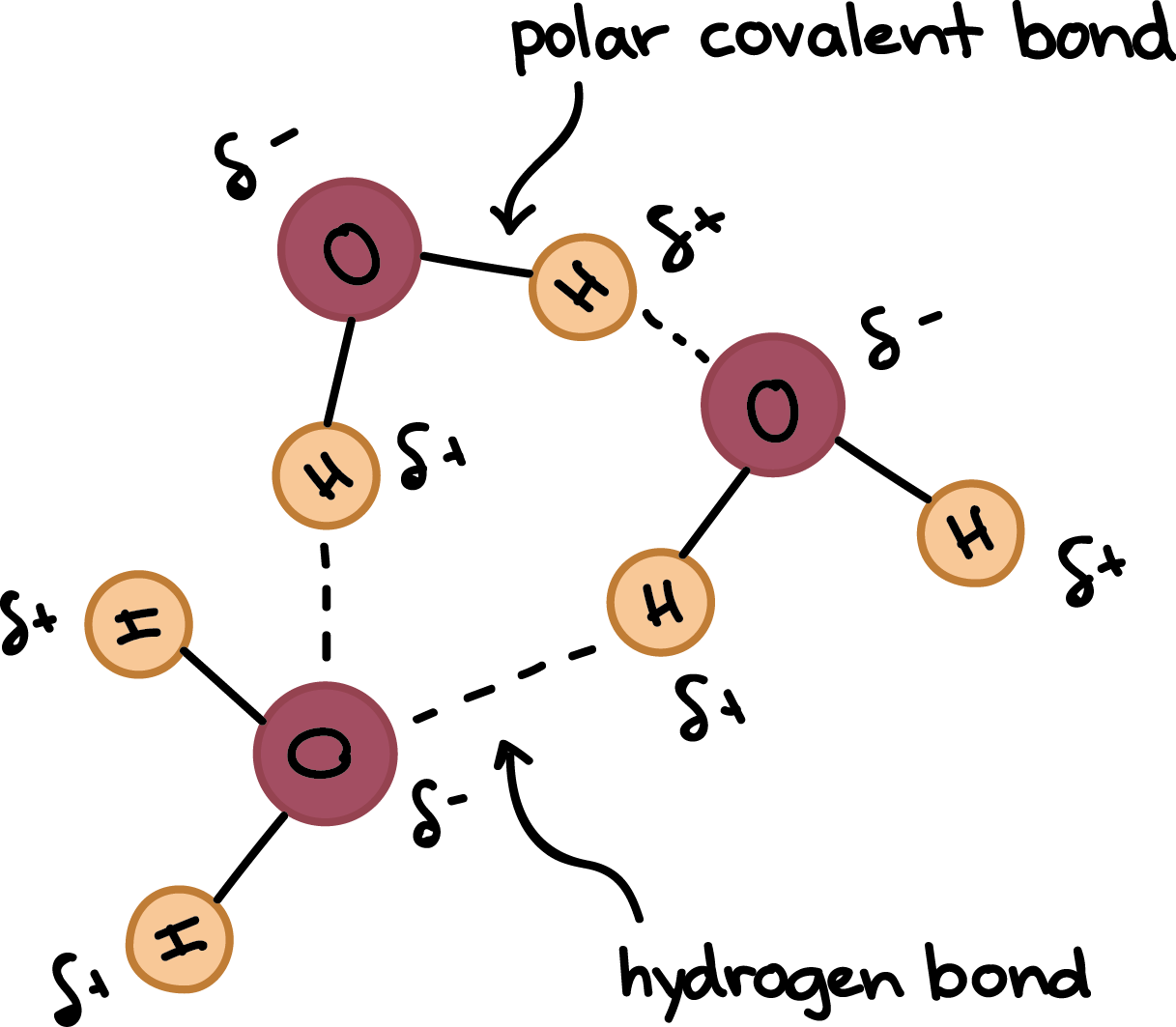
📸 Source/Description: Polarity and hydrogen bonding in water
B. Properties as a Solvent
Water is called the "universal solvent" because its polarity allows it to dissolve other polar molecules and ionic compounds, forming hydration shells.
C. Importance in Biological Systems
- Medium for Life: It is the primary constituent of cells where biochemical reactions occur.
- Transport Medium: Carries nutrients, gases, and waste products (e.g., in blood).
- Temperature Regulation: Has a high specific heat capacity and high heat of vaporization, helping organisms maintain a stable internal temperature.
- Reactant/Product: Participates directly in hydrolysis and dehydration synthesis reactions.
- Structural Role: Contributes to turgor pressure in plants and helps maintain the shape of macromolecules.
VII. Key Takeaways for IMAT Preparation
For success in the IMAT biology section concerning "The Chemistry of Living Things," focus on the following core concepts:
- Structure-Function Relationships: Understand how a molecule's structure dictates its function (e.g., α vs. β linkages in carbohydrates; R-groups in amino acids).
- Monomers and Polymers: Know the building blocks for each class (monosaccharides, amino acids, nucleotides) and the bonds that link them (glycosidic, peptide, phosphodiester bonds).
- Key Examples and Their Roles: Be familiar with glucose, glycogen, cellulose, phospholipids, cholesterol, DNA, and RNA.
- Comparisons and Contrasts: Differentiate between related molecules (e.g., starch vs. cellulose; DNA vs. RNA).
- Water's Centrality: Appreciate how water's polarity and hydrogen bonding give rise to its life-sustaining properties.
Developing an appreciation for these interrelationships will provide a more holistic and integrated understanding of cellular chemistry, which is invaluable for medical studies.