
Genetics and Reproduction
An Advanced Review for Pre-Medical Students
Table of Contents>
I. Mendelian Genetics Basics
This section covers the fundamental principles of heredity as discovered by Gregor Mendel, which form the bedrock of classical genetics. Understanding these laws is essential for predicting patterns of inheritance.
A. Key Terminology
| Term | Definition |
|---|---|
| Gene | A specific sequence of nucleotides in DNA that is located on a chromosome and that is the functional unit of inheritance. |
| Allele | One of two or more alternative forms of a gene that arise by mutation and are found at the same place on a chromosome. |
| Locus | The specific physical location of a gene on a chromosome. |
| Genotype | The genetic makeup of an organism, represented by the combination of alleles it possesses (e.g., TT, Tt, tt). |
| Phenotype | The observable physical or biochemical characteristics of an organism, determined by its genotype and environmental factors (e.g., tall, short). |
| Homozygous | Having two identical alleles for a particular gene (e.g., TT or tt). |
| Heterozygous | Having two different alleles for a particular gene (e.g., Tt). |
| Dominant Allele | An allele that expresses its phenotypic effect even when heterozygous with a recessive allele (e.g., T). |
| Recessive Allele | An allele that only expresses its phenotypic effect when two copies are present (homozygous) (e.g., t). |
Homologous Chromosomes with Alleles
Image SourceB. Mendel's Laws
| Law | Core Principle | Associated Cross | Key Ratios (F2 of heterozygote cross) |
|---|---|---|---|
| Law of Segregation | During gamete formation (meiosis), the two alleles for a heritable character separate from each other so that each gamete ends up with only one allele. | Monohybrid Cross (examines one trait) | Phenotypic: 3:1, Genotypic: 1:2:1 |
| Law of Independent Assortment | Alleles of genes on non-homologous chromosomes assort independently during gamete formation. The inheritance of one character has no effect on the inheritance of another. | Dihybrid Cross (examines two traits) | Phenotypic: 9:3:3:1 |
Punnett Square for Monohybrid Cross (Tt x Tt)
Image SourcePunnett Square for Dihybrid Cross (RrYy x RrYy)
Image SourceC. Non-Mendelian Inheritance
Many traits do not follow simple Mendelian patterns. These exceptions provide a more nuanced understanding of genetics.
| Pattern | Description | Example |
|---|---|---|
| Incomplete Dominance | The heterozygous phenotype is an intermediate blend of the two homozygous phenotypes. | Red (CRCR) and white (CWCW) snapdragons produce pink (CRCW) offspring. |
| Codominance | Both alleles are fully and separately expressed in the heterozygous phenotype. | Human ABO blood group system, where alleles IA and IB are both expressed in type AB blood. |

Incomplete Dominance in Snapdragon Flowers
Image Source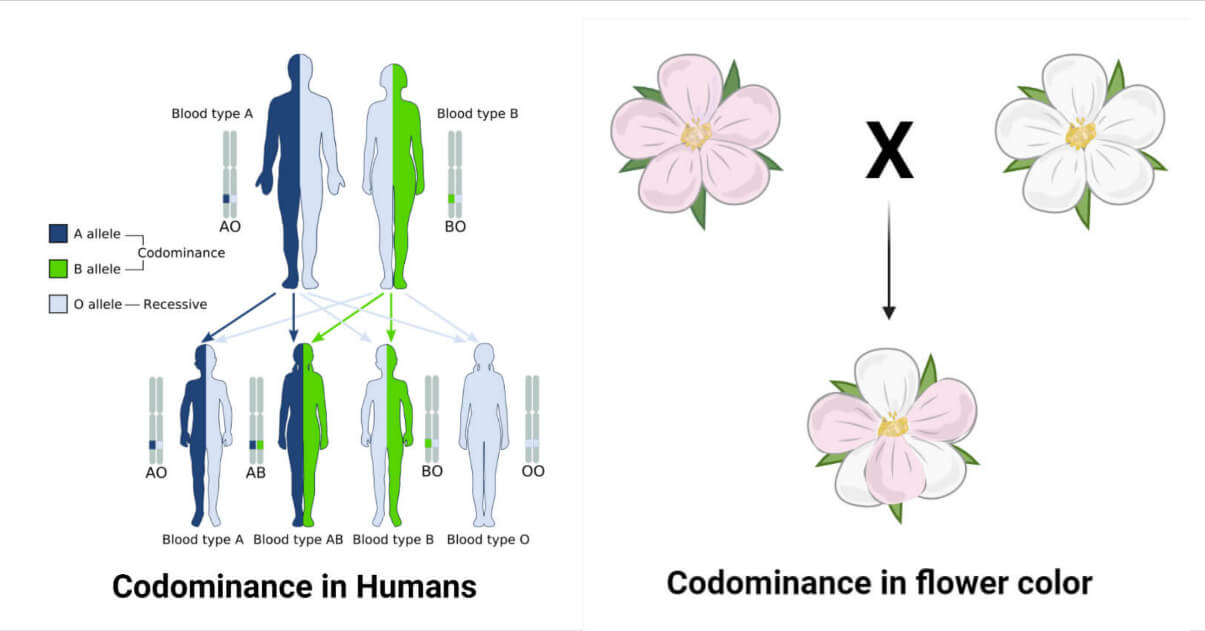
Codominance in Human ABO Blood Types
Image SourceD. Human Genetics Tools
Analyzing inheritance in humans requires specialized tools due to ethical and practical constraints on experimental breeding.
| Tool | Description & Use |
|---|---|
| Pedigree Chart | A family tree diagram that shows the inheritance of a trait over several generations. Used to determine the mode of inheritance (autosomal dominant/recessive, X-linked) and to calculate probabilities of inheriting a trait. |
| Karyotype | An organized profile of a person's chromosomes. Used to diagnose chromosomal abnormalities, such as aneuploidy (e.g., Trisomy 21 - Down Syndrome) or large structural changes. |
Example of a Pedigree Chart (Autosomal Dominant)
Image SourceHuman Karyotype
Image SourceII. Molecular Genetics
This section explores the molecular mechanisms that underpin heredity: the structure and function of DNA, and how the genetic code is expressed as functional proteins.
A. DNA Replication
Before a cell divides, it must accurately duplicate its entire genome. This process, known as DNA replication, is semiconservative and relies on the coordinated action of several key enzymes.
| Enzyme/Protein | Function in DNA Replication |
|---|---|
| Helicase | Unwinds the DNA double helix at the replication fork. |
| Single-Strand Binding Proteins (SSBs) | Bind to and stabilize single-stranded DNA, preventing it from re-pairing. |
| Topoisomerase | Relieves the torsional strain ahead of the replication fork caused by unwinding. |
| Primase | Synthesizes a short RNA primer, providing a 3'-OH end for DNA polymerase to start synthesis. |
| DNA Polymerase III | (In prokaryotes) The main replication enzyme, synthesizing the new DNA strand by adding nucleotides to the 3' end. |
| DNA Polymerase I | (In prokaryotes) Removes the RNA primers and replaces them with DNA nucleotides. |
| DNA Ligase | Joins the Okazaki fragments on the lagging strand to create a continuous DNA strand. |
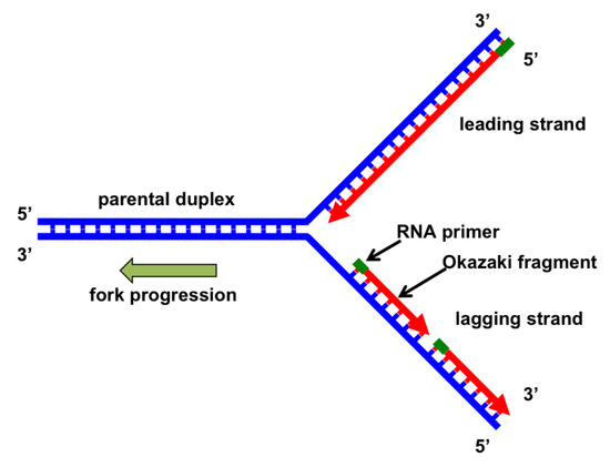
DNA Replication Fork
Image SourceB. From Gene to Protein: The Central Dogma
The central dogma of molecular biology, proposed by Francis Crick, describes the two-step process, transcription and translation, by which the information in genes flows into proteins: DNA → RNA → Protein.
1. Transcription
This is the synthesis of an RNA copy of a segment of DNA. In eukaryotes, this process occurs in the nucleus and involves several steps, including post-transcriptional modification of the primary transcript (pre-mRNA) into mature mRNA.
Transcription Process
Image Source2. Translation
This is the synthesis of a polypeptide, using the information in the mRNA. The process occurs on ribosomes in the cytoplasm. The mRNA sequence is read in codons (groups of three bases), and tRNA molecules bring the corresponding amino acids to the ribosome.
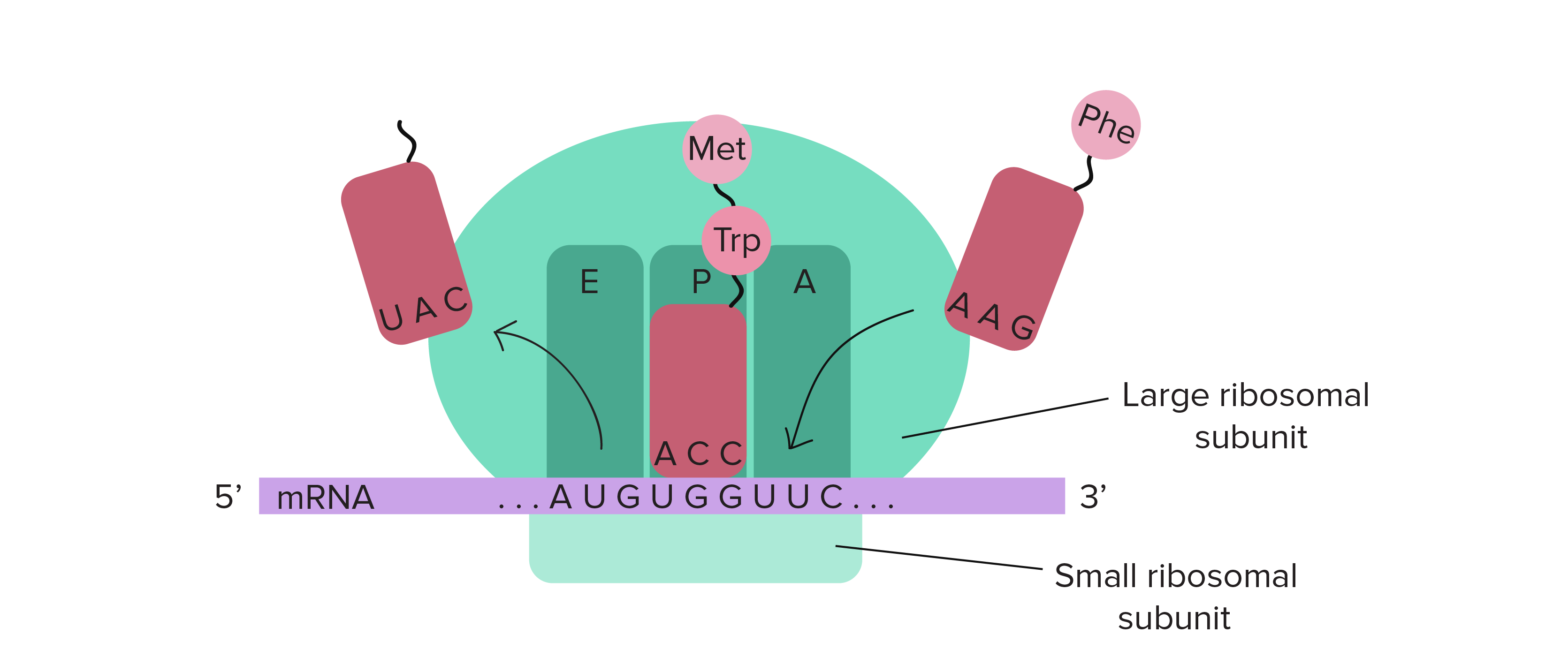
Translation at the Ribosome
Image SourceC. Mutations
A mutation is a permanent alteration in the DNA sequence. They are the ultimate source of genetic variation but can also be the cause of genetic diseases.
| Types of Gene (Point) Mutations | |
|---|---|
| Substitution (Silent) | A base change that results in a codon that codes for the same amino acid. No effect on the protein. |
| Substitution (Missense) | A base change that results in a codon that codes for a different amino acid. Can have minor or major effects (e.g., sickle cell anemia). |
| Substitution (Nonsense) | A base change that results in a premature stop codon. Leads to a truncated, usually nonfunctional, protein. |
| Insertion/Deletion (Frameshift) | Addition or loss of nucleotides (not in a multiple of three) that shifts the reading frame, altering all subsequent amino acids and often creating a premature stop codon. |

Gene Mutations (Point Mutations)
Image SourceChromosomal Mutations
Image SourceIII. Genetic Technology
The understanding of molecular genetics has led to powerful technologies that allow us to analyze and manipulate DNA.
| Technique | Principle and Key Applications |
|---|---|
| Recombinant DNA Technology |
|
| Polymerase Chain Reaction (PCR) |
|
| CRISPR-Cas9 Gene Editing |
|
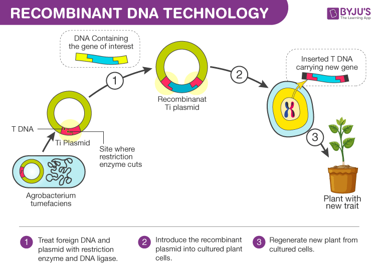
Recombinant DNA Technology Flowchart
Image Source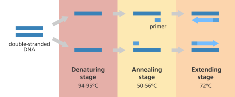
Polymerase Chain Reaction (PCR) Cycle
Image SourceCRISPR-Cas9 Gene Editing Mechanism
Image SourceIV. Eukaryotic Gene Regulation
Unlike prokaryotes, eukaryotic gene expression is regulated at multiple levels, allowing for complex control of cell differentiation and function. This intricate system ensures that the right genes are expressed at the right time and in the right place.
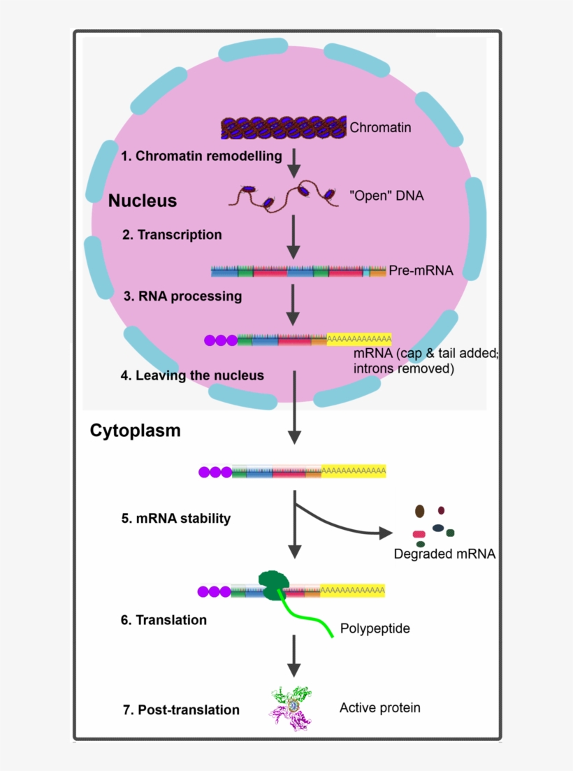
Eukaryotic Gene Expression Regulation
Image Source| Level of Regulation | Mechanism | Example/Supplement |
|---|---|---|
| Pre-Transcriptional | Control of DNA accessibility through chromatin modification. Tightly packed heterochromatin is generally silent, while loosely packed euchromatin is transcriptionally active. |
|
| Transcriptional | The primary control point, involving the binding of transcription factors to specific DNA sequences to initiate or block transcription. |
|
| Post-Transcriptional | Modification and processing of the primary RNA transcript (pre-mRNA) in the nucleus before it is exported to the cytoplasm. |
|
| Translational | Regulation of the rate at which mRNA is translated into protein. |
|
| Post-Translational | Modification of the polypeptide chain after synthesis to make it a functional protein. |
|
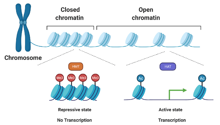
Histone Acetylation and Chromatin Remodeling
Image SourceV. Advanced Molecular Processes
A deeper look into the eukaryotic-specific mechanisms of DNA replication reveals key differences from the prokaryotic model, particularly in managing linear chromosomes and ensuring high fidelity.
Eukaryotic DNA Replication Details
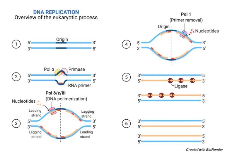
Eukaryotic Replication Fork with Polymerase Switching
Image Source| Feature | Description in Eukaryotes |
|---|---|
| Multiple Origins of Replication | To replicate the vast eukaryotic genome in a timely manner, DNA synthesis is initiated at many points (origins) simultaneously. These are recognized by the Origin Recognition Complex (ORC). |
| Specialized DNA Polymerases | Eukaryotes use a larger, more complex set of DNA polymerases with distinct roles:
|
| The End-Replication Problem & Telomerase | DNA polymerase cannot fully replicate the 3' end of the lagging strand, leading to progressive shortening of linear chromosomes with each cell division. The enzyme Telomerase, a reverse transcriptase with its own RNA template, extends the repetitive DNA sequences at the ends (telomeres), counteracting this shortening in germline cells, stem cells, and many cancer cells. |
Action of Telomerase
Image SourceVI. Gene Mutations & Molecular Basis of Disease
Specific types of mutations at the DNA level can have profound clinical consequences, leading to a wide range of genetic disorders by altering protein structure and function.
| Mutation Type | Molecular Consequence | Classic Disease Example |
|---|---|---|
| Missense | A single nucleotide substitution results in a codon that codes for a different amino acid. The effect can range from benign to severe depending on the new amino acid's properties and location. | Sickle Cell Anemia: A single A-to-T substitution in the β-globin gene replaces the hydrophilic Glutamic acid (Glu) with the hydrophobic Valine (Val), causing hemoglobin to polymerize under low oxygen. |
| Nonsense | A nucleotide substitution results in a premature STOP codon (UAA, UAG, UGA), leading to the synthesis of a truncated, and typically non-functional, protein. | Duchenne Muscular Dystrophy: Many cases are caused by a nonsense mutation in the dystrophin gene, which is crucial for muscle fiber integrity. The resulting truncated protein leads to progressive muscle wasting. |
| Frameshift | An insertion or deletion of a number of nucleotides that is not a multiple of three. This alters the triplet reading frame of the codons, usually leading to a completely different downstream amino acid sequence and often a premature stop codon. | Tay-Sachs Disease: A common cause is a 4-base insertion in the HEXA gene. This frameshift results in a non-functional hexosaminidase A enzyme, causing a fatal buildup of lipids in brain cells. |
| Splice-Site | A mutation at an intron-exon boundary (splice donor or acceptor site) disrupts the normal splicing of pre-mRNA. This can lead to the retention of an intron or the exclusion of an exon in the mature mRNA, altering the protein product. | β-Thalassemia: Certain mutations in the HBB gene occur at splice sites, leading to incorrect splicing of the β-globin pre-mRNA. This results in a deficient production of functional hemoglobin, causing severe anemia. |
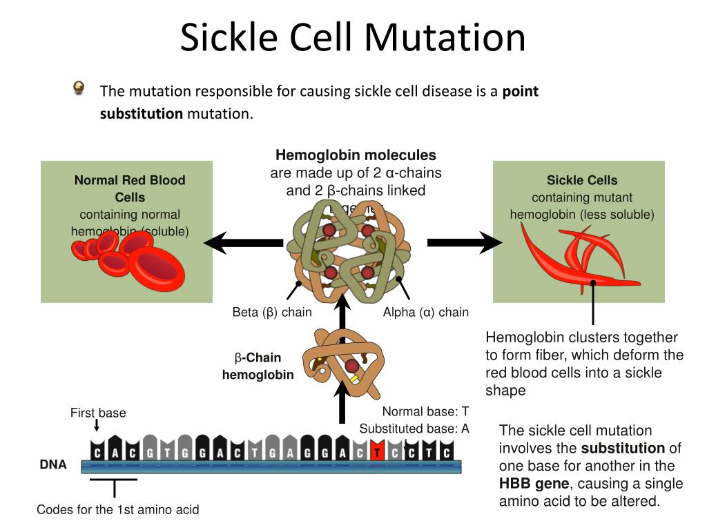
Sickle Cell Anemia Mutation
Image Source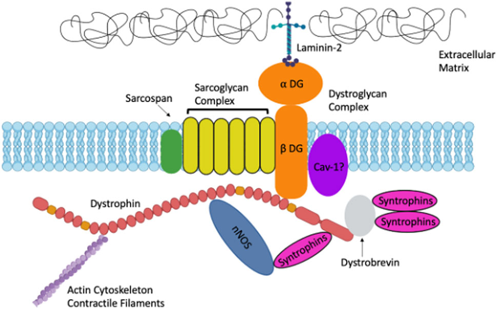
Effect of Dystrophin Mutation in DMD
Image SourceVII. Chromosomal Abnormalities & Diagnostics
Large-scale changes at the chromosomal level, involving number or structure, can also cause significant genetic syndromes. Various cytogenetic and molecular techniques are used for their diagnosis.
| Abnormality Type | Description | Example | Diagnostic Techniques |
|---|---|---|---|
| Numerical (Aneuploidy) | Gain or loss of one or more entire chromosomes, typically resulting from non-disjunction during meiosis I or II. |
| Karyotyping: Visualizes full chromosome set. FISH: Uses fluorescent probes for specific chromosomes. |
| Structural | Alterations affecting parts of a chromosome, such as deletions, duplications, inversions, or translocations (exchange of segments between non-homologous chromosomes). |
| Karyotyping: Detects large structural changes. Array CGH: Detects small deletions/duplications (microdeletions/microduplications) with high resolution. |
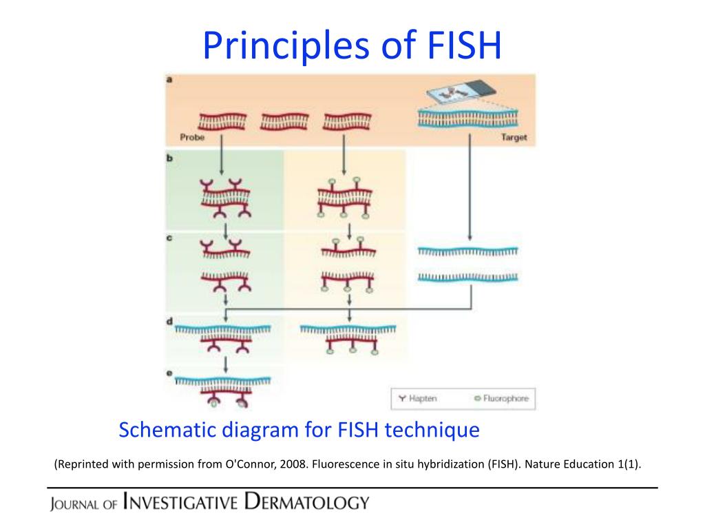
Fluorescence In Situ Hybridization (FISH)
Image Source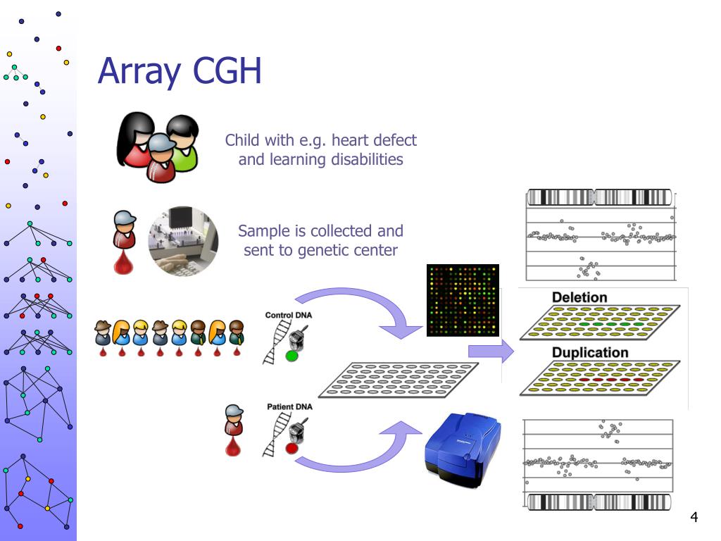
Array CGH (aCGH) Workflow
Image SourceVIII. Advanced Genetic Technologies & Applications
Beyond the foundational techniques, several advanced methods are crucial in modern research and diagnostics, enabling personalized medicine and deeper biological insights.
| Technique | Principle | Primary Application |
|---|---|---|
| Quantitative PCR (qPCR) | Also known as real-time PCR, this technique uses fluorescent dyes or probes to measure the amount of amplified DNA in real-time during a PCR reaction. The rate of fluorescence increase is proportional to the initial amount of target DNA. | Gene expression analysis (by reverse transcribing mRNA to cDNA first), viral load quantification (e.g., HIV, COVID-19), and pathogen detection. |
| RNA Sequencing (RNA-Seq) | A high-throughput, next-generation sequencing (NGS) method that captures a snapshot of the entire transcriptome (all RNA molecules) in a cell or tissue. It provides information on which genes are expressed and at what levels. | Comprehensive gene expression profiling, discovering novel genes and transcript isoforms, identifying biomarkers for diseases like cancer, and understanding cellular responses to stimuli. |
| SNP Analysis & GWAS | Detecting Single Nucleotide Polymorphisms (SNPs), which are the most common type of genetic variation. Genome-Wide Association Studies (GWAS) scan the genomes of many individuals to find SNPs associated with a particular trait or disease. | Identifying genetic risk factors for complex diseases (e.g., diabetes, heart disease), predicting individual drug responses (pharmacogenomics), and ancestry tracing. |
Quantitative PCR (qPCR) Amplification Plot
Image Source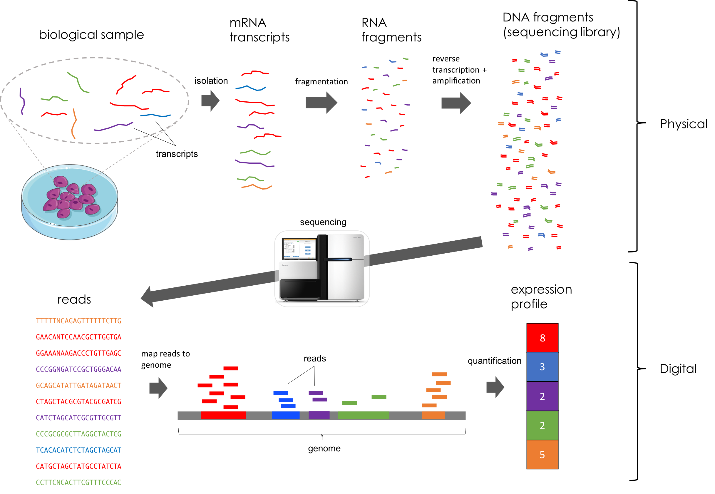
RNA-Sequencing (RNA-Seq) Workflow
Image Source