An exhaustive, university-level masterclass exploring Osteology, Bone Histology, Ossification Processes, Joint Classifications, and Calcium Homeostasis dynamics.
Est. Reading Time: 210 Mins 100% Curriculum Sync 30-Question Simulator
Lecture 1: Functions & Macroscopic Anatomy of Bone
The human skeleton is not merely a dry, inert scaffold; it is a dynamic, living organ system. It provides structural support, facilitates movement via leverage, protects vital internal organs, acts as a massive mineral reservoir, and serves as the exclusive site for hematopoiesis (blood cell formation).
Bone is a specialized form of dense connective tissue with a mineralized matrix, providing both rigidity and metabolic flexibility.
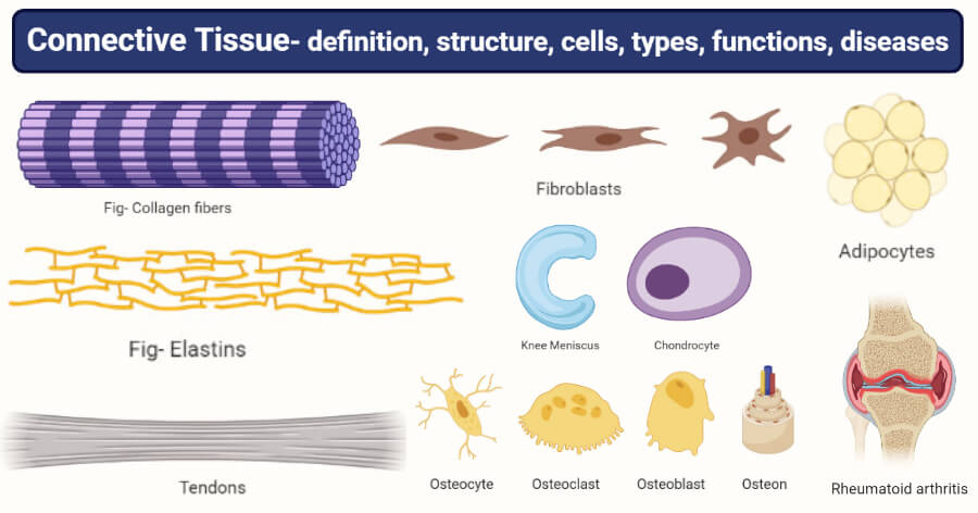
Figure 1.1: Histological classification of connective tissues, including bone.
1.1 Division of the Skeleton
The adult human skeleton consists of 206 named bones, structurally divided into two major groups based on their location and primary functional role.
The Axial Skeleton
Forms the long central axis of the body. Its primary function is the rigid protection, support, and carrying of other body parts.
The Skull (Cranium & Facial bones)
The Vertebral Column
The Bony Thorax (Ribs & Sternum)
The Appendicular Skeleton
Consists of the bones of the upper and lower limbs and the girdles that attach them to the axial skeleton. Its primary function is locomotion and environmental manipulation.
Upper & Lower Limbs
Pectoral (Shoulder) Girdle
Pelvic (Hip) Girdle
1.2 Classification of Bones by Shape
Bones are classified into four distinct categories based on their gross morphological shape, which intimately dictates their structural function.
1. Long Bones
Considerably longer than they are wide. Composed of a central shaft (diaphysis) and two distinct, expanded ends (epiphyses). Almost all bones of the limbs (e.g., Femur, Humerus, Phalanges) fall into this category.
2. Short Bones
Roughly cube-shaped. They contain mostly spongy bone covered by a thin layer of compact bone. Examples include the carpals (wrist) and tarsals (ankle). A special subtype, Sesamoid bones (e.g., the patella), form entirely within tendons.
3. Flat Bones
Thin, flattened, and usually slightly curved. Composed of two parallel layers of compact bone sandwiching a layer of spongy bone (diploe). Examples include the Sternum, Scapulae, Ribs, and most cranial bones.
4. Irregular Bones
Bones that exhibit complex, convoluted shapes that do not fit the other categories. Consist of spongy bone enclosed by thin layers of compact bone. Classic examples are the Vertebrae and the Coxal (hip) bones.
1.3 Macroscopic Structure of a Typical Long Bone
Figure 1.0: Gross anatomy of a generic long bone.
The Diaphysis (Shaft)
Forms the long axis of the bone. It is constructed of a thick collar of highly dense Compact Bone that completely surrounds a central cavity. In adults, this central cavity is called the Medullary Cavity and contains fat (Yellow Marrow).
The Epiphyses (Ends)
The expanded ends of the bone. The exterior is composed of a thin layer of compact bone, while the interior houses a honeycomb-like network of Spongy (Cancellous) Bone. The joint surfaces of epiphyses are covered with friction-reducing Articular (Hyaline) Cartilage.
The Epiphyseal Line separates the diaphysis and epiphysis. It is a remnant of the Epiphyseal Plate, the disc of hyaline cartilage that actively grows during childhood to lengthen the bone.
1.4 Bone Membranes: Periosteum & Endosteum
The Periosteum
A glistening white, double-layered membrane that covers the entire external surface of the bone (except joint surfaces). The outer fibrous layer is dense irregular connective tissue. The inner osteogenic layer contains stem cells (osteoprogenitors), osteoblasts, and osteoclasts. It is richly supplied with nerve fibers and massive blood vessels, which enter the diaphysis via nutrient foramina. It is anchored to the bone by strong collagen tufts called Sharpey's fibers.
The Endosteum
A delicate connective tissue membrane covering all internal bone surfaces. It covers the trabeculae of spongy bone and lines the canals passing through compact bone. Like the inner periosteum, it contains osteogenic stem cells.
Lecture 2: Bone Histology & Cellular Architecture
Bone (osseous tissue) is a specialized form of connective tissue. It is incredibly hard, yet remarkably lightweight. Its unique properties arise from its composite matrix—a perfect synergy of organic protein fibers providing flexibility and tensile strength, heavily impregnated with inorganic mineral salts providing immense compressional hardness.
2.1 The Cells of Bone Tissue
Five major cell types populate bone tissue, working in concert to continuously build, maintain, and remodel the matrix.
1. Osteoprogenitor (Osteogenic) Cells
Mitotically active stem cells located in the periosteum and endosteum. When stimulated, they differentiate rapidly into osteoblasts. Some remain as stem cells to maintain the pool.
2. Osteoblasts (The Builders)
Bone-forming cells. They actively secrete the unmineralized organic bone matrix called Osteoid (which consists primarily of collagen and calcium-binding proteins). They are highly actively mitotic when building matrix.
3. Osteocytes (The Maintainers)
Mature bone cells completely trapped within small spaces called Lacunae in the solid matrix they created. They are no longer mitotic. They monitor and maintain the bone matrix and act as stress/strain sensors to direct remodeling.
4. Osteoclasts (The Destroyers)
Giant, multinucleate cells derived from the same hematopoietic stem cell lineage as macrophages (NOT from osteoprogenitors). They reside in shallow depressions called resorption bays. They possess a ruffled border that heavily secretes lysosomal enzymes and brutal acid to rapidly digest and resorb bone matrix.
2.2 Chemical Composition of Bone
The remarkable durability of bone is due to its composite nature, balancing flexibility with extreme hardness.
Organic Components (35%)
Includes the cells and the Osteoid. Osteoid consists of ground substance and massive amounts of Collagen fibers. The collagen is structured in specific, alternating alignments.
Function: Provides immense tensile strength and flexibility, allowing bone to twist and bend slightly without snapping.
Inorganic Components (65%)
Consists almost entirely of Hydroxyapatites (mineral salts), primarily complex calcium phosphates. These tiny mineral crystals are tightly packed in and around the collagen fibers of the extracellular matrix.
Function: Provides the defining characteristic of bone—extreme hardness and resistance to brutal compressional forces.
2.3 Microscopic Anatomy of Compact Bone (The Osteon)
Compact bone appears incredibly dense and solid to the naked eye, but under a microscope, it is riddled with canals acting as passageways for nerves and massive blood vessels. The structural unit of compact bone is the Osteon (or Haversian System).
Figure 2.0: Transverse section of a single Osteon. The osteocytes are trapped in solid rock, but remain connected to the blood supply and each other via the spider-web network of canaliculi.
Anatomy of the Osteon
An osteon is an elongated cylinder oriented parallel to the long axis of the bone. Functionally, it acts like a microscopic weight-bearing pillar.
Concentric Lamellae:
Each osteon is a group of hollow tubes of bone matrix placed tightly inside one another, much like the rings of a tree trunk. Each individual tube is a lamella. Crucially, the collagen fibers in adjacent lamellae run in entirely opposite directions. This alternating pattern acts as a brilliant 'twister resister', allowing the bone to withstand immense torsional (twisting) stress.
The Central (Haversian) Canal:
Runs straight through the core of each osteon. It contains small blood vessels and nerve fibers serving the needs of the osteon's cells.
Lacunae & Canaliculi:
Spider-shaped osteocytes occupy small hollow spaces (Lacunae) situated strictly at the junctions of the lamellae. Hair-like canals called Canaliculi radiate aggressively from the lacunae, connecting them to each other and directly to the central canal. This massive network ties all the osteocytes together, allowing nutrients and wastes to be actively passed from cell to cell via gap junctions throughout the solid bone cylinder.
Perforating (Volkmann's) Canals:
Lie at right angles to the long axis. They brutally connect the blood and nerve supply of the periosteum to those in the central canals and the deep medullary cavity.
2.4 Microscopic Anatomy of Spongy Bone
Unlike compact bone, spongy bone completely lacks true osteons. Instead, it consists of a haphazard, honeycomb-like network of tiny bone struts called Trabeculae.
These trabeculae appear incredibly disorganized, but they are actually aligned precisely along lines of physical stress, acting like flying buttresses to brace the bone. They contain irregularly arranged lamellae and osteocytes connected by canaliculi. There are no central canals; nutrients diffuse directly into the trabeculae from the rich red or yellow marrow filling the open spaces between them.
Lecture 3: Osteogenesis (Bone Development & Growth)
Osteogenesis (ossification) is the complex process of bone tissue formation. In human embryos, before week 8, the skeleton is constructed entirely from soft, flexible fibrous connective tissue membranes and massive chunks of hyaline cartilage. These soft tissues act as a blueprint. The replacement of this soft blueprint with hard bone occurs via two entirely distinct pathways.
3.1 Intramembranous Ossification
Bone develops directly from a fibrous connective tissue membrane, without a cartilage intermediate.
The Process:
Mesenchymal (stem) cells cluster within the membrane and differentiate directly into osteoblasts. These osteoblasts immediately begin secreting osteoid (matrix). The matrix quickly calcifies, trapping the cells (which become osteocytes). Blood vessels aggressively invade the area, and woven spongy bone forms around them. Finally, compact bone replaces the outer spongy bone layers.
Bones Formed: Primarily flat bones (Cranial bones of the skull and the Clavicles).
3.2 Endochondral Ossification
Bone forms by the replacement of a Hyaline Cartilage model. This process is much more complex because the existing cartilage must be systematically destroyed as the bone is laid down.
The Sequential Stages:
A bone collar forms solidly around the diaphysis of the hyaline cartilage model.
Cartilage specifically in the center of the diaphysis rapidly calcifies and then develops massive cavities (as the cartilage cells choke and die).
The Periosteal Bud (a massive complex of blood vessels, nerves, osteoblasts, and osteoclasts) violently invades these internal cavities and forms the primary spongy bone. This is the Primary Ossification Center.
The diaphysis violently elongates and a massive medullary cavity forms as osteoclasts smash the new spongy bone. Shortly before or after birth, Secondary Ossification Centers appear directly in the epiphyses (ends).
The epiphyses ossify completely. When complete, hyaline cartilage remains in only two places: on the epiphyseal surfaces (articular cartilage) and at the junction of the diaphysis and epiphysis, forming the crucial Epiphyseal Plates.
Bones Formed: Essentially all bones below the base of the skull (except clavicles).
3.3 Postnatal Growth: Elongation vs. Appositional
Longitudinal Growth (Length)
Occurs exclusively at the Epiphyseal Plate. Cartilage cells continuously undergo rapid mitosis on the epiphyseal side (pushing the end away), while simultaneously being destroyed and replaced by bone on the diaphyseal side. This keeps the plate a constant thickness while the bone aggressively lengthens.
At the end of adolescence, under the influence of surging sex hormones, the cartilage cells halt mitosis. The entire plate is rapidly replaced by solid bone (Epiphyseal Plate Closure), permanently halting longitudinal growth.
Appositional Growth (Width)
Growing bones must massively widen as they lengthen to prevent buckling. Osteoblasts located beneath the periosteum secrete new bone matrix onto the external bone surface. Simultaneously, osteoclasts on the endosteal surface aggressively smash and remove bone.
Normally, the builders outpace the destroyers slightly, producing a thicker, significantly stronger bone without making it impossibly heavy.
Lecture 4: Bone Remodeling & Calcium Homeostasis
Bone is incredibly dynamic. Every week, roughly 5-7% of our total bone mass is recycled. The entire adult skeleton is completely replaced every 10 years. This continuous process of bone deposition and bone resorption is known as Bone Remodeling, orchestrated entirely by osteoblasts and osteoclasts.
4.1 The Dual Control System
Remodeling is not random; it is strictly controlled by two incredibly powerful, distinct physiological loops.
1. Mechanical Stress (Wolff's Law)
Determines where remodeling occurs. Wolff's law simply states that a bone aggressively grows or radically remodels entirely in response to the physical demands or mechanical stresses placed upon it.
Long bones are incredibly thick precisely midway along the diaphysis, exactly where bending stresses are maximal.
Curved bones are thickest exactly where they are most likely to buckle.
Trabeculae of spongy bone form perfect structural trusses aligned exactly along lines of physical compression.
Massive, heavy projections occur exclusively where massive, active skeletal muscles attach and pull violently.
2. Hormonal Control ($Ca^{2+}$)
Determines whether and when remodeling occurs. The body physically needs calcium for muscle contraction, nerve transmission, and blood coagulation. If systemic blood calcium levels drop, the body will ruthlessly cannibalize the skeleton to get it.
Parathyroid Hormone (PTH): When blood $Ca^{2+}$ drops, the parathyroid glands dump PTH. PTH rapidly signals aggressive Osteoclasts to massively degrade the bone matrix, releasing huge volumes of $Ca^{2+}$ directly into the blood, ignoring the structural integrity of the bone.
A joint is any site where two or more bones physically meet. Their fundamental role is to give the rigid skeleton mobility while holding it together tightly. Joints are classified both functionally (by how much they move) and structurally (by what material binds them).
5.1 Functional vs. Structural Classification
Structural Class (Binding Material)
Functional Class (Mobility)
Characteristics & Prime Examples
Fibrous
Synarthroses
(Immovable)
Bones are joined rigidly by dense fibrous connective tissue. No joint cavity is present.
Sutures: Tight seams between cranial bones.
Syndesmoses: Connected exclusively by ligaments (e.g., distal tibiofibular joint).
Gomphoses: The specific "peg-in-socket" fibrous joint holding a tooth securely in its alveolar socket.
Cartilaginous
Amphiarthroses
(Slightly Movable)
Bones are intimately united by cartilage. No joint cavity is present.
Synchondroses: A bar or plate of hyaline cartilage (e.g., Epiphyseal plates, 1st rib to sternum). Mostly immovable.
Symphyses: Articular surfaces covered with hyaline cartilage are subsequently fused to an intervening pad of compressible fibrocartilage (e.g., Intervertebral discs, Pubic symphysis). Acts as a heavy shock absorber.
Synovial
Diarthroses
(Freely Movable)
The articulating bones are entirely separated by a fluid-filled joint cavity. This highly complex arrangement permits vast freedom of movement. All joints of the limbs fall into this ultimate structural class.
5.2 Architecture of a Synovial Joint
Synovial joints are complex biophysical machines. They all share six distinguishing architectural features.
1. Articular Cartilage
Glassy-smooth hyaline cartilage covers opposing bone surfaces. Absorbs massive compression to keep bone ends from being crushed.
2. Joint (Articular) Cavity
A unique potential space completely filled with a tiny amount of synovial fluid.
3. Articular Capsule
Encloses the cavity. Two layers: Outer tough Fibrous Layer (dense irregular CT) to prevent bones pulling apart. Inner Synovial Membrane (loose CT) which actively manufactures synovial fluid.
4. Synovial Fluid
A viscous, incredibly slippery fluid derived from blood. Contains hyaluronic acid. Weeping lubrication prevents the immense friction that would aggressively overheat and destroy joint tissues.
5. Reinforcing Ligaments
Massive, thickened bands of dense regular CT that cross the joint to limit excessive or dangerous ranges of motion.
6. Nerves and Blood Vessels
Richly supplied with sensory nerve fibers monitoring pain and rapidly relaying joint position/stretch to the cerebellum. Extensive capillary beds in the synovial membrane aggressively produce the synovial fluid.
Clinical Pathology: Arthritis
Osteoarthritis (OA)
The 'wear-and-tear' arthritis. Degenerative. Years of intense mechanical stress physically degrade the articular cartilage faster than it can be replaced. Exposed bone thickens and forms bony spurs (osteophytes) restricting movement.
Rheumatoid Arthritis (RA)
A brutal, chronic inflammatory autoimmune disease. The immune system violently attacks its own synovial membranes. The inflamed membrane thickens into a destructive Pannus that physically erodes the articular cartilage and underlying bone, eventually fusing the joint solid (ankylosis).
Lecture 6: The Comprehensive IMAT Skeletal Database
For the IMAT examination, rote memorization of histological architecture, ossification pathways, and specific axial/appendicular bones is non-negotiable. The following exhaustive table compiles the entirety of the skeletal system covered in the masterclass into an ultra-high-yield, rapid-review format.
Structure / Cell / Process
Anatomical/Histological Location
Primary Physiological Function & Distinguishing Features
Bone Cells
Osteoprogenitors
Periosteum/Endosteum. Mitotic stem cells; differentiate into osteoblasts.
Osteoblasts
Actively building surfaces. Synthesize and intensely secrete organic Osteoid (collagen matrix).
Osteocytes
Trapped within solid Lacunae. Maintain the existing matrix. Act as vital stress sensors connecting via Canaliculi.
Osteoclasts
Resorption bays. Giant multinucleate cells derived from macrophages. Aggressively dissolve bone using severe acid and lysosomal enzymes. Triggered massively by PTH.
Rings of bone matrix. Alternating collagen direction provides massive resistance to extreme twisting (torsion) forces.
Central (Haversian) Canal
Runs longitudinally. Houses the primary blood vessels and nerve fibers serving the individual osteon.
Perforating (Volkmann's) Canals
Run transversely. Connect the blood supply of the periosteum deeply into the central canals and medullary cavity.
Canaliculi
Microscopic hair-like canals linking all lacunae together, allowing gap junction communication through solid rock.
Ossification Processes
Intramembranous Ossification
Bone develops directly from fibrous connective tissue membrane. Forms the flat bones of the skull and clavicles.
Endochondral Ossification
Bone aggressively replaces a hyaline cartilage model. Requires destruction of cartilage and invasion by a periosteal bud. Forms almost all long bones.
Structural Joint Classes
Fibrous (Synarthroses)
Bones fused rigidly by dense fibrous CT. No cavity. (e.g., Cranial Sutures).
Cartilaginous (Amphiarthroses)
Bones fused by cartilage. No cavity. (e.g., Symphyses like Intervertebral discs acting as massive shock absorbers).
Synovial (Diarthroses)
Fluid-filled joint cavity separating bones. Articular cartilage, synovial membrane, and ligaments permit extreme, free movement. (e.g., All limb joints).
Part 7: The IMAT Skeletal Simulator
This massive, comprehensive 30-question examination rigorously tests the exhaustive details presented in all preceding lectures of this masterclass. Designed strictly at the official IMAT difficulty level, it focuses heavily on histological differentiation, ossification pathways, clinical joint pathology, and strict calcium homeostasis. Do not begin until you have absolutely mastered the material above.